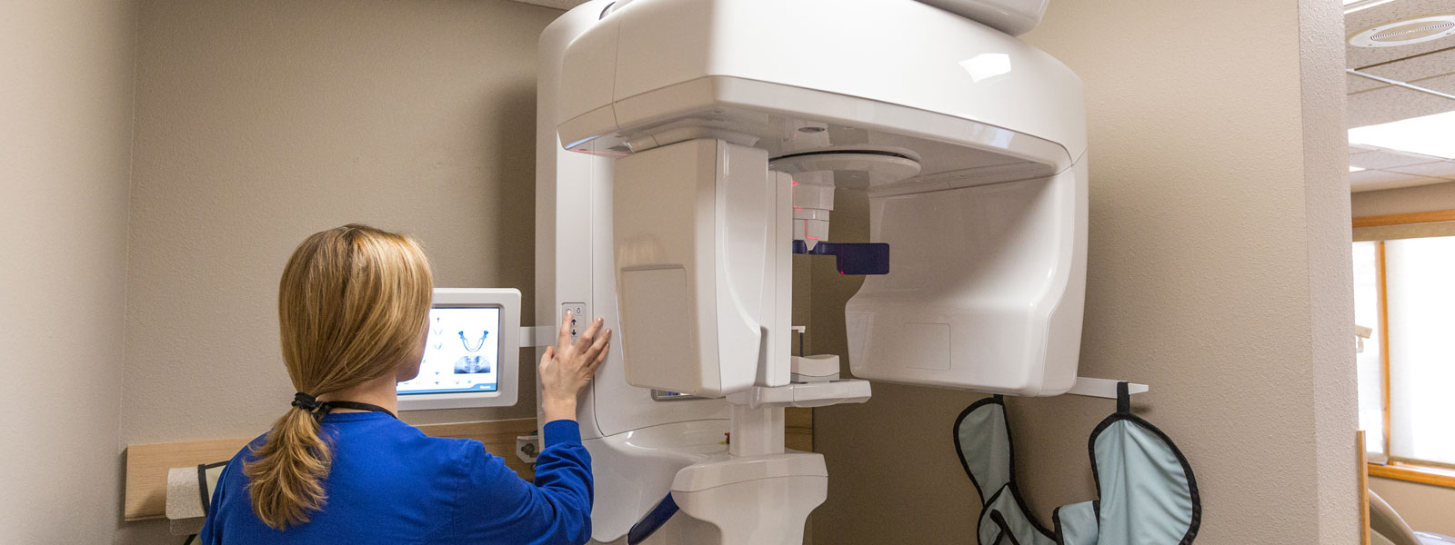
In December of 2014 the practice purchased a cone beam x-ray unit. Cone beam imaging is relatively new technology to dentistry but is having a profound effect on the way dentists can now gather information. Cone beam scans use a cone-shaped beam that is directed to a detector that rotates around the patient. The cone beam images are converted into three-dimensional images, giving the dentist the ability to see all structures in any given field. Highly complex software takes these hundreds of slices of information and reconstructs them according to the needs of the dentist, giving him precisely the application needed to diagnose and treatment plan. Oral anatomy not visible with traditional two-dimensional images is now revealed in clarity with cone beam imaging.
Dr. Smith is now placing dental implants, and this new technology is a powerful tool in analyzing if you would be a good candidate for an implant, and in accurate placement once the treatment is undertaken.
After many wonderful years, Dr. Jonathan Smith is retiring. We are proud to continue caring for you with the same team, in the same location, under the trusted leadership of Dr. Trason Shoquist, DMD.
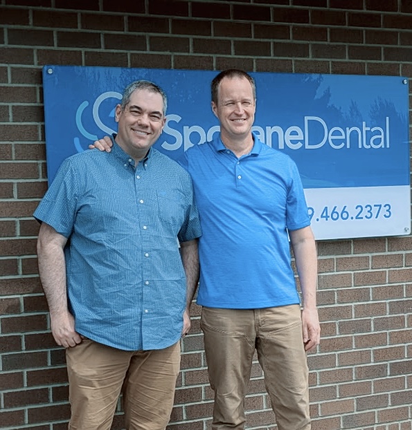
Dr. Trason Shoquist, DMD
U.S. Army veteran · Roseman University · A patient-first approach rooted in listening.