What is Cone Beam Dental Imaging?
At Spokane Dental we have the highest standard of care and health for our patients, including state of the art imaging. Dental Cone Beam Imaging is relatively new technology in the dental field that provides unparalleled 3D imaging of not only a patient’s teeth, but their jaw and sinuses as well. We use this imaging for many aspects of care for our patients, and it is a key aspect in the high quality care we offer every patient that walks through the Spokane Dental doors.
What is Cone Beam Imaging?
Cone Beam imagining uses a special type of technology differing from standard dental x-rays that provides a 3 dimensional image. Dr. Smith is not only able to see the crown and root of the tooth and the jaw, but nerve paths and soft tissue as well.
Traditional dental x-rays show only hard tissue in 2D: bone and tooth, essentially. There is very important vasculature and soft tissue that can be detected through cone beam imaging that just isn’t visible with traditional x-rays. What is Getting Cone Beam Imaging Like?
What is Getting Cone Beam Imaging Like?
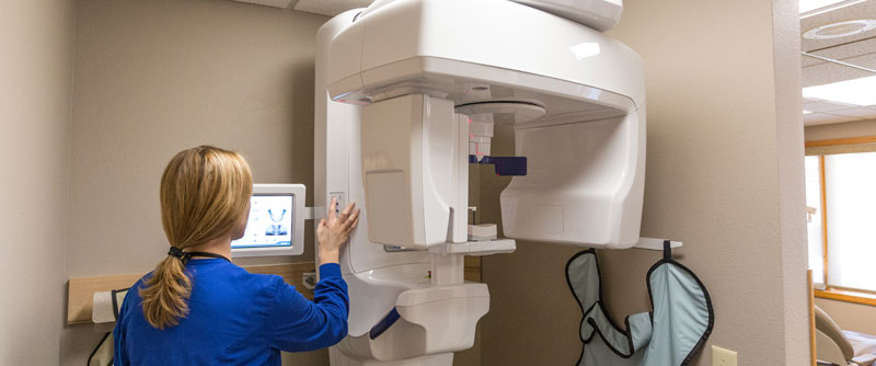
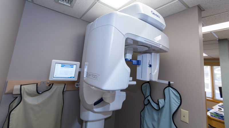
Cone Beam Imaging in our office is simple, straightforward, and fast. The chin rest and bite stick line up the patient perfectly for the imaging. While the patient stands with their head aligned properly (chin in chin rest, forehead resting on guide, and gently biting on stick to ensure teeth are spaced properly), the small arms of the machine move in a circle around the patient, capturing images.
The machine takes many “pictures” from many different angles, which are all put together to make the 3D image of the patient’s anatomy. It is completely digital and instantly available for Dr. Smith to read.
Why Do I Need This Imaging?
Dr. Smith uses cone beam imaging both for diagnosing oral disease or problems as well as treatment planning procedures. There are several ways that dental Cone Beam Imaging is useful in the dental office:
Dental Implants:
Dental Implants involve replacing a tooth with an implant that acts like a tooth: root and all. A post is implanted into the jaw (with a crown eventually on top that looks like a real tooth), allowing the patient normal use of the implant as if it were a tooth. Cone Beam Imaging is the gold standard for x-rays and imaging when it comes to dental implants.
With the imaging we have in our practice, Dr. Smith can plan and then place implants in the safest, most effective way possible. Dr. Smith can ensure that the angle the implant is placed allows for the healthiest bone levels and growth surrounding the implant, and that no nerves are compromised.
Primary and Permanent Dentition:
Permanent Dentition, or “adult teeth” can be seen on dental cone beam imaging during formation and before they even erupt. On a child, a cone beam image can show the primary dentition or “baby teeth”, as well as the permanent teeth that are getting ready to erupt. This is an excellent diagnostic tool for children to ensure that all of the adult teeth are forming how they should, as well as will be able to erupt in the correct time frame.
Diagnosing Disease:
Many oral diseases, maladies, or problems can be diagnosed through a dental cone beam image. Screening for certain oral cancers (such as jaw tumors) can be done through this type of imaging, as well as TMJ Disorder, screening of the sinuses, impacted wisdom teeth, and much more.
No matter the dental concern, your team at Spokane Dental is here to help! Led by Dr. Smith, our hygienists, assistants, and office staff are friendly and here to provide the best possible health for our patients. Whether you are in need of a routine cleaning, interested in dental implants, or anything in between we are just a phone call away. Reach us at: (509)466-2373.
Blog Post Author

Karissa Barker BSDH | Contributor
Spokane Dental